OUR CASE
Matt was taken from ED to MRI and then straight to OT for decompression and stabilisation of his C-Spine.
He comes to ICU intubated and ventilated with an art line but no CVC for ongoing management…
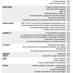
Click below for a brief checklist of things to think about on an ICU ward round for a patient with an acute SCI.
Feel free to print it off.
A BIT MORE DETAIL…
SURGERY
Spinal cord compression may occur due to misalignment of spine, oedema or haemorrhage.
Incomplete injury with spinal cord compression should be urgently decompressed.
Stability is the ability of the spine, under physiological loads, to limit patterns of displacement, which would result in neurological injury, pain, or deformity. It is affected by fracture morphology and osteo-ligamentous integrity.
Most unstable spinal injuries require operative intervention.
Where indicated, early surgery (within 8 hours of injury) is safe, may improve neurological outcome, reduces length of stay in acute care, permits early mobilization and commencement of DVT prophylaxis and lowers hospital cost REF
Most spinal surgeons prefer to operate as early as possible in patients with incomplete or cervical spinal cord injury.
DRUGS
Many pharmacological agents have been investigated secondary injury and aid recovery of damaged neurons; however none of these have proven successful.
NASCIS 2 and NASCIS 3 were the largest trials that investigated the effects of corticosteroids in SCI.
They both were negative studies, showing no significant difference in motor score between treatment groups at any time points.
Post-hoc sub-group analysis of this data suggesting neurological outcome benefit was fundamentally flawed.
The lack of evidence of efficacy and the adverse effects of steroids such as sepsis (particularly pneumonia), detrimental effects on concomitant head injury, risk of gastrointestinal bleeding and pancreatitis, has seen steroid use fall out of favour in recent years.
Other agents that have been investigated with no evidence of benefit include GM-1 ganglioside, tirilazad, naloxone and nimodipine.
BLOOD PRESSURE
There is weak evidence that a mean arterial pressure (MAP) > 85 mmHg should be maintained for 5- 7 days following injury, theoretically improving cord perfusion pressure and preventing secondary injury.
Common practice is After initial fluid resuscitation vasopressors such as noradrenaline are commonly required to meet this target.
Like with surgery, this makes the most sense in incomplete injuries – it’s hard to imagine in a completely transected cord that hypertensive therapy can make a difference.
There are no published randomised controlled trials on this practice. Therefore it cannot yet be considered established best practice, and the risks of such therapy need careful consideration on a case by case basis.
The duration of hypertensive therapy becomes more contentious when you have otherwise well paraplegic patients confined to ICU for pressor support, which may be delaying early spinal rehab
TEMPERATURE
Induced hypothermia has been reported to be beneficial in case reports but there is not enough evidence to recommend this intervention currently.
Fever may worsen outcome and normothermia should be maintained.
WHY IT’S DIFFERENT
> In cervical and upper thoracic SCI the diaphragm provides 90% of the tidal volume. The resting position of the diaphragm can make a big difference
> Secretions are increased and often thicker
> Cough is affected so secretion clearance affected
> Patients may experience “air hunger” feeling like they’re not getting enough tidal volume, from loss of innervation of stretch receptors
AN APPROACH
In the initial period after cervical SCI, ventilator settings are dictated by associated injuries and other co-morbidities.
For example, in patients with severe traumatic brain injury, normocapnia should be maintained.
Once acute issues have resolved, mechanical ventilation should be tailored to SCI.
Baseline spirometry should be obtained to allow monitoring of progress.
The literature on ventilatory management of acute SCI is very limited and of low quality.
Ventilator tidal volumes (VT) in excess of 10 ml/kg are often used in patients with cervical SCI.
The rationale for high VT ventilation is to prevent atelectasis and the common sensation of “air hunger” experienced following SCI.
This avoids the need for high level PEEP that may lower the position of diaphragm to one of mechanical disadvantage.
In some spinal centers VT of 10-20 ml/kg has been used routinely.
With high VT, dead-space must be incorporated into the circuit to avoid hypocapnoea.
Another approach is to use more conventional VT and higher PEEP to prevent atelectasis. Until better quality evidence is available, an individualised approach to ventilator settings should be used.
One approach to mechanical ventilation for patients with “healthy” lungs:
> Use an assisted, pressure-controlled, ventilation mode
> Adjusted to achieve tidal volumes of 10 to 12 mL/kg
> 5 to 7 cm H2O of PEEP
> Plateau pressure below 30 cm H2O.
The goal is to maintain total ventilator support, allowing the patient to initiate most of the cycles and attempting to adjust the inspiratory time to adapt it to the neural inspiratory time.
When ready, introduce short trials of spontaneous respiration with rest periods.
The trial periods should start shorter than with other ventilatory wean patients to avoid exhausting patients e.g. 5 minutes per hour
EXTUBATION
Extubation after mechanical ventilation in patients with cervical SCI requires careful consideration of several factors including:
> No imminent procedures or imaging planned
> Gas exchange adequate
> Reversible pathology treated (e.g. infection, pulmonary oedema, pleural collections, sputum plugs)
> Assessed for ability to tolerate non-invasive ventilation
> Secretion load manageable
> Vital Capacity > 10 ml/kg
> Reintubation considered
Patients may benefit from extubation to non-invasive ventilation directly, which may be required for several days post extubation.
A variety of non-invasive ventilation interface devices should be available and nasal skin should be protected from developing pressure ulcers.
Aggressive 24/7 chest physiotherapy and assisted coughing are often required initially to clear secretions.
TRACHEOSTOMY
Failure of extubation usually means a tracheostomy.
Extubation failure is commonly due to retained, excessive secretions, inadequate cough and sputum plugs.
The tracheostomy rate after cervical SCI varies between 20-60%.
Complete injuries above C5 usually require tracheostomy.
A high NLOI, complete SCI, associated facial fractures, thoracic trauma and emergency intubation are associated with need for tracheostomy.
Percutaneous tracheostomy is as safe as surgical tracheostomy.
Early tracheostomy may reduce the duration of mechanical ventilation and ICU length of stay and it does not increase the risk of wound or implant infection even after anterior spinal fixation.
WEANING
Weaning from MV after cervical SCI may take considerable time.
In some centers, high-level tetraplegics are cared for in chronic respiratory care facilities rather than ICU.
It is important that a long term weaning strategy is agreed upon and adhered to by all members of the multidisciplinary team.
Accurate neurological assessment, progressive ventilator free breathing guided by vital capacity, adequate rest periods with controlled ventilation, and secretion control with aggressive physiotherapy are associated with successful weaning.
Inspiratory muscle training uses an inspiratory resistor to increase strength of inspiratory muscles.
While more studies are needed to clearly delineate the role of inspiratory muscle training in weaning of patients with cervical SCI, its use makes physiological sense and is being used increasingly.
It should be remembered that most patients with cervical SCI ventilate more easily when supine compared to being upright, as their abdominal contents can push the diaphragm up into a position of better mechanical advantage in supine position.
An abdominal binder may be used in the sitting position, which also helps in this regard.
Inhaled anticholinergics and beta-2 agonists reduce airway resistance but have not been shown to improve success of weaning.
Anabolic steroids like oxandrolone do not lead to significant improvement in respiratory function and may cause hepatotoxicity and dyslipidemia.
Phrenic nerve or direct diaphragmatic pacing systems are occasionally used for respiratory support in high-level tetraplegics, more here.
The NLOI is the strongest predictor of successful weaning.
Patients with complete injuries at or above C3 are likely to need permanent ventilation.
Early identification of patients who cannot be weaned helps reduces their ICU length of stay and expedites the arrangements for equipment and care personnel required for home ventilation or transfer to a chronic respiratory care facility.
Only 2% of patients with SCI require life-long ventilation. Most of these have complete injures at C1-C4.
NEUROGENIC SHOCK
A distributive type of shock seen in SCI
From loss of segmental sympathetic innervation causing decreased vasomotor tone / systemic vascular resitance in periperal vasculature
Causes pooling of blood in peripheries, decreased venous return
Associated bradycardia in cervical SCI as heart loses sympathetic innervation from the upper thoracic spine but parasympathetic supply from vagus is not affected
Decreased venous return and decreased heart rate reduces cardiac output
> Leads to secondary SCI, organ dysfunction and death if not treated
> Occurs in 24-43% of cervical SCI and 19% of thoracic SCI REF
Other causes of hypotension, particularly hypovolemic shock must be excluded before a diagnosis of neurogenic shock is made.
Bradycardia and even asystole may occur during routine ICU procedures like endotracheal suction or repositioning in bed in patients with a cervical NLOI.
This phenomenon occurs most commonly in the first week and generally resolves within two to six weeks after injury.
MANAGEMENT
> Judicious IV fluid resuscitation initially – small aliquots with regular reassessment of volume status
> Invasive arterial blood pressure monitoring
> Central venous catheter insertion, subclavian or PICC ideal, avoid IJ if collar in situ, avoid femoral as increased risk of VTE
> Commence vasopressors for MAP > 85 mmHg
- Noradrenaline infusion as default
- If very bradycardic consider adrenaline, or isoprenaline.
- If just bradycardic and only for short periods copnsider glycopyyrollate or atropine
- If no central access consider metaraminol infusion
Common
Without adequate prophylaxis incidence up to 100%
With prophylaxis still 5%
PE in top causes of mortality REF
LMWH superior to unfractionated heparin in reducing DVT but not PE and carries less bleeding complications
AN APPROACH
> Surgery as early as possible
> Start enoxaparin 40 mg daily in < 72 hours from injury (unless contraindication)
> If anticoagulation contraindicated consider vena caval filter
> Screening lower limb doppler ultrasound weekly in ICU
ILEUS
Paralytic ileus and acute gastric dilatation are common initially following SCI.
The upper motor neuron bowel syndrome seen following lesions above the cauda equina; it is characterised by increased colonic wall and anal tone with loss of external anal sphincter control, which results in constipation and faecal retention.
Defaecation may be induced by a reflex activity caused by a rectal stimulus such as an irritant suppository or digital stimulation.
Other components of bowel management programs include:
> Adequate fluid intake
> Normal diet
> Laxatives (usually docusate and macrogol)
> Daily suppositories commonly required (polyethylene glycol-based)
> Avoid opiates and other constipating medications
> Very rarely, surgery or electrical stimulation.
In lower motor neuron bowel syndrome, seen in cauda equina injuries, the external anal sphincter is lax but colonic peristalsis is inhibited leading to both constipation and overflow incontinence.
STRESS ULCERS
Unopposed parasympathetic activity results in stomach hyperacidity
> Use a proton pump inhibitor for ulcer prophylaxis
CHRONIC PAIN
Frequent complication after SCI that occurs in up to two-thirds of patients.
Pain after SCI may be classified as nociceptive pain from stimulation of nociceptors; or neuropathic pain from damage to sensory system itself.
Most patients report onset of chronic pain within six months after the injury, and frequently whilst still in ICU.
WHICH DRUGS WORK?
Gabapentinoids (gabapentin and pregabalin) for neuropathic pain have the strongest evidence to support their efficacy.
Tricyclic antidepressants such as amitriptyline are effective for neuropathic pain in patients who also have depression.
Infusions of opiates, ketamine and lignocaine are effective for neuropathic pain in the short term.
Tramadol reduces neuropathic pain in SCI and there is evidence for tapentadol ‘s efficacy in other neuropathic pain syndromes.
Cannabinoids, intrathecal baclofen, and botulinum toxin remain the most supported treatments for nociceptive or spasticity-related pain.
Behavioral therapy may be useful in addition to pharmacological treatment.
Consultation with a pain specialist should be sought in patients with severe, refractory pain.
REFERENCE
AN APPROACH
> Start with paracetamol
> Limit opioids to acute perioperative period
> If pain has neuropathic qualities commence gabapentin and trial tapentadol
> If concomitant depression add amitriptyline
> If neuropathic pain persists, obtain pain team consult
Most patients with a NLOI above L1 experience detrusor over activity associated with detrusor sphincter dyssynergia.
This results from the upper motor neuron lesion causing over-activity of both detrusor and bladder sphincters. As a result, the detrusor contracts against a closed sphincter, leading to high intravesical pressure, vesico-uretheral reflux, high residual volume, incontinence, bladder spasm and an increased risk of urinary tract infections.
Most patients with complete cervical SCI will require a supra pubic cystostomy, while thoracolumbar injuries may be managed by intermittent self-catheterisation.
Bladder spasticity is common and anticholinergics agents like oxybutynin, solifenacin, tolterodine or trospium should be commenced early in the admission to prevent a contracted, low-volume bladder.
The anticholinergic side effects of these agents may be significant, including constipation. Some patients require a combination of two anticholinergic medications.
Following SCI, there is a marked alteration in calcium homeostasis, with potential hypercalcaemia, hypercalciuria and a significant risk of calcium oxalate nephrolithiasis.
APPROACH
> Commence anticholinergic agent as soon as bowels are opened e.g. solifenacin 5 mg daily
> Obtain baseline CT of the renal tract c. 2 months following SCI
> Suspect renal calculi and urinary tract infections as precipitants of autonomic dysreflexia
Pressure ulcers are common due to immobility and lack of sensation.
10-30% of patients with SCI develop pressure ulcers.
They are a source of significant morbidity and are associated with increased hospital length of stay, life threatening infections, chronic refractory osteomyelitis and autonomic dysreflexia.
Skin healing in denervated skin is significantly delayed, so prevention is vital.
The incidence of pressure areas may be reduced by:
- Daily skin inspection to detect early pressure areas
- Prevention of accumulation of moisture
- Good bowel and bladder care
- Pressure relieving mattresses
- Protocol-based frequent patient repositioning
- Optimal nutrition
- Patient participation in their skin care program

At day 5 ICU Matt has been extubated and is progressing well.
He’s being hypertensed to a MAP of > 85 mmHg for 5 days as per local protocol.
His wound drain was only removed on day 4 and the surgeons involved are keen to keep him off anticoagulation this week for fear it may affect wound healing.
What do you reckon?

