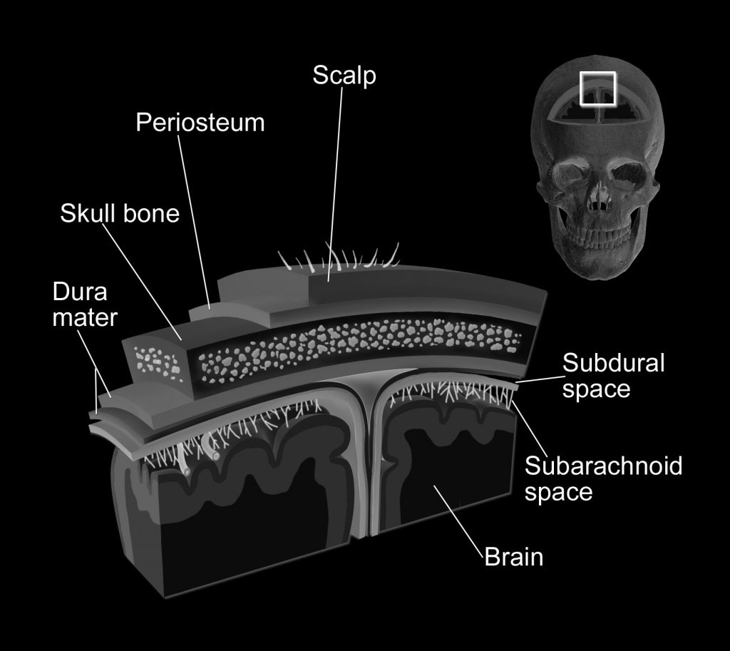
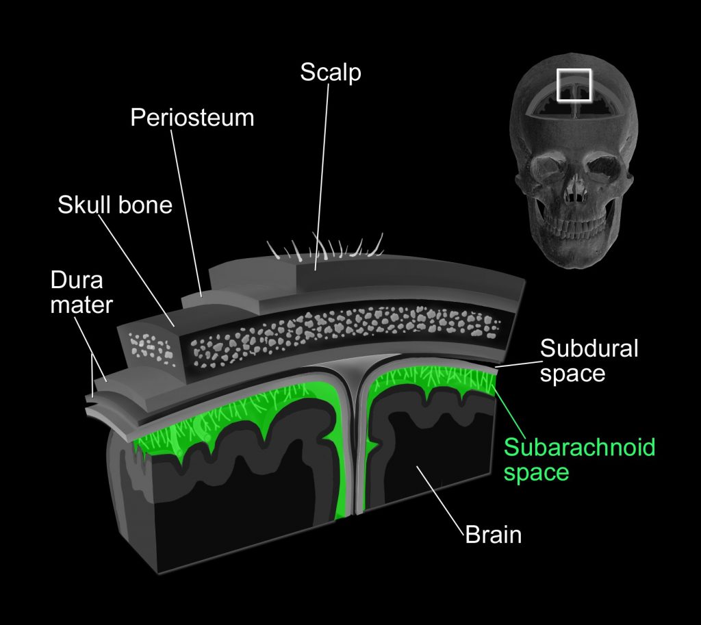
Definition
A subarachnoid haemorrhage is bleeding into the subarachnoid space
VASCULAR ANATOMY
Before we go any further, you will need to have a general understanding of the arterial supply to the brain.
The brain is supplied by the
> Left and right internal carotid arteries
> Left and right vertebral arteries
These arteries unite within the skull (through communicating arteries) creating the “Circle of Willis”, supplying much of the brain.
For those needing a 2 minute refresher on the blood supply to the brain please watch the following video as it will held understand some of the finer points to come. Otherwise keep moving along to the CT angiograms below!
PATHOPHYSIOLOGY OF SAH BRAIN INJURY
Image summarising pathophysiology EBI and DCI
Coming soon
Coming soon
Coming soon
Coming soon
Coming soon
Coming soon
> Reactive microglial response
Oxyhaemoglobin
Microthrombi in glymphatics
Touch the different vessels on this circle of Willis to see what’s what in this CT Angiogram
Scroll through this spin around the cerebral vessels to appreciate how it works in 3 dimensions
EPIDEMIOLOGY
Incidence
2-20 per 100,000 of population.
More common in Japan & Finland
Less common in Central & South America
Gender
F : M = 1.6 : 1
Age
Peak 45-64 years
RISK FACTORS


MODIFIABLE
Hypertension
Cigarette smoking
Alcohol abuse
Sympathomimetic drugs such as cocaine
NOT pregnancy, delivery or puerperium
AETIOLOGY
What causes aneurysms?
The exact cause of intracranial aneurysms are incompletely understood, though may be:
Congenital
- Defect in the muscular layer of the arterial wall, referred to as a medial gap
Atherosclerotic or hypertensive.
- The presumed etiology of most berry aneurysms and probably interacts with congenital predisposition.
Infectious
- So called “mycotic aneurysms”, making up <5% of all intracranial aneurysms.
- Most commonly found in MCA territory (80% of time) and caused by staph and strep spp. (65%)
- Commonly occur in patients with bacterial endocarditis and the immunocompromised
Traumatic
- <1% intracranial aneuryms; more commonly pseudoaneuryms
- Commonly caused by closed head injuries; less commonly iatrogenic / open head injuries.
Embolic
- Rare cause, predominately MCA territory
- 10–15% of patients with atrial myxoma develop metastatic aneurysms
Where do aneurysms commonly occur?
~85% IN CAROTID SYSTEM
30% Anterior Communicating Artery (AComA)
25% Posterior Communicating Artery (PComA)
20% Middle Cerebral Artery (MCA)
10% Other locations within carotid system
~15% IN POSTERIOR CIRCULATION
10% on basilar artery:
- Basilar artery bifurcation (basilar tip) is the most common
- Others: Basilar-Superior Cerebellar artery (BA-SCA) junction, Basilar-Vertebral artery (BA-VA) junction, anterior inferior cerebellar artery (AICA)
5% on vertebral artery:
- Vertebral artery-posterior inferior cerebellar artery (VA-PICA) junction is the most common
MULTIPLE
25% of aneurysm patients have multiple aneurysms
Other causes of spontaneous SAH
Most common “other cause”
- Cerebral arteriovenous malformation (note, these more commonly cause ICH & IVH)
Less common “other causes”
- Vasculitis
- Cerebral artery dissection
- Coagulation disorders (pathological or medication related)
- Dural sinus thrombosis
Rare “other causes”
- Rupture of an infundibulum
- Tumors
- Pre-truncal nonaneurysmal SAH
- Drugs including cocaine
- Sickle cell disease
- Pituitary apoplexy


